

What Type Of Aortic Anuerysm Is Easiest To Repair
Blazon Iv Thoracoabdominal Aneurysm Repair
![]() Indications/Contraindications
Indications/Contraindications
Indications
Thoracoabdominal aneurysms (TAA) develop through a circuitous process involving a combination of genetic and ecology factors. Approximately 80% are degenerative, 15% to twenty% are secondary to chronic dissection and the remaining have other etiologies such as infection or aortitis. TAA are classified co-ordinate to the scheme originally devised by Crawford (Fig. sixteen.1). This classification is specially useful in patients requiring operative repair since it has straight implications for both the technical deport of the operation, and the incidence of operative complications; in particular, ischemic spinal cord injury. This chapter will focus on Blazon Iv TAA which accept an upper limit of the crus of the diaphragm (or 13th intercostal infinite) and can extend to the iliac bifurcation.
The critical business with TAA is rupture, which is associated with a mortality that approaches xc% and accounts for thirty% of all deaths in patients with TAA. The strongest predictor of rupture is aneurysm size. Natural history studies have shown that the hazard of rupture is negligible in TAA less than five cm, equivalent to the risks associated with open surgery in the 5 to half dozen cm range, and increases substantially when the diameter exceeds 6 cm and/or the charge per unit of interval growth is ≥10 mm per yr. These observations have led to the acceptance of vi cm as a generally appropriate size threshold for recommendation of surgical intervention for Type Iv TAA. Other run a risk factors for rupture include increasing patient age, female person gender, pain (including atypical pain), a history of chronic obstructive pulmonary illness (COPD), symptoms attributable to the aneurysm, and dissection developing within a degenerative aneurysm. Population-based studies have indicated an overall 5-year rupture risk of 20%, with lxxx% of ruptures occurring in women.
Contraindications
Patient option for open repair of Type Four TAA is predicated upon weighing the likelihood of aneurysm rupture against the run a risk of surgery as conferred by the patient'southward medical history and agile comorbidities. An accurate assessment of associated comorbid conditions is mandatory to guide advisable decision making with respect to recommending open surgical
repair. Patients with agile cardiac affliction, capacity-limiting COPD, and renal failure are at highest gamble for postoperative complications and decease after open up TAA repair. These are patients in whom a nonstandard open (hybrid) repair or no intervention may be warranted.

Figure 16.1 Crawford classification of thoracoabdominal aneurysms.
![]() Preoperative Planning
Preoperative Planning
Medical Evaluation
All patients should be evaluated with dipyridamole thallium scanning or the equivalent thereof to assess perioperative myocardial ischemic potential. In addition, patients with a history of symptoms suggestive of heart failure should take an assessment of left ventricular office. While patients with significant impairments of pulmonary reserve can usually be detected on a historical ground alone, nosotros routinely obtain preoperative pulmonary function studies. Advanced historic period is an of import component only in as much as it is accompanied by overall fragility and dumb functional status (both of which have been shown to predict mortality).
Imaging
Associated vascular diseases and comorbid conditions are commonplace in patients presenting for treatment of thoracic aneurysms. Synchronous aneurysms typically involving the ascending aorta or arch have been observed in some x% of patients being evaluated for descending thoracic aneurysm repair. Prior performance for aortic aneurysm disease is seen in one-tertiary of patients and as noted above, the about common of these is a previous infrarenal aneurysm repair. Additionally, up to one-quarter of patients with thoracic aortic affliction take concomitant abdominal aortic aneurysm (AAA), therefore all patients should undergo baseline imaging of the abdominal and pelvic aorta at the time of initial diagnosis. In contemporary practice, a dynamic, fine-cutting, contrast-enhanced CT browse with or without helical reconstruction is the preferred imaging modality and provides: (1) Accurate cess of aneurysm size and extent, (ii) a baseline report to which futurity images may be compared, and (iii) important anatomic information such every bit the anatomy and topography of the visceral segment, relationship of the left renal vein to the aorta (anterior or posterior) and appropriate areas for clamping (Fig. 16.ii).
Accurate assessment of the size of Blazon Four TAA is contingent upon measurement in the appropriate perpendicular airplane. Three-dimensional reconstruction of the thoracic aorta with decision of the centerline of flow ensures that whatsoever cantankerous-sectional measurement will be perpendicular to the aortic axis. The quality of current CT scanners with three-dimensional
reconstruction is exceptional and in our practice, this has become the prototype modality of choice for the evaluation and handling of thoracic aortic aneurysms (Fig. 16.3).
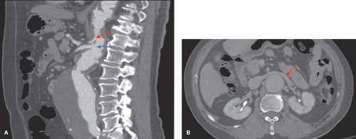
Effigy sixteen.2 A: Lateral reconstruction of the aorta shows the relationship of the origin of the celiac artery (red pointer) to the SMA (blue arrow). B: Axial image of the visceral aorta. The red arrow points to the anterior left renal vein which is displaced past the aneurysm.
![]() Surgery
Surgery
Principles of Handling
Our general approach to the repair of Blazon IV TAA is the clamp-and-sew technique. Hypothermic renal protection is usually employed. A bolus (250 cc) of renal preservation
fluid (4°C lactated Ringers with 25 g of mannitol per liter and 1 thousand methyl prednisolone per liter) is infused directly into the ostia of each renal avenue after the aorta is opened. This is followed past a continuous drip of the aforementioned delivered through vi-Fr perfusion balloon-tipped catheters. The initial bolus results in a decline of renal parenchymal temperature to 15°C and the subsequent continuous infusion maintains the renal core temperature effectually 25°C equally measured past direct temperature probes in the renal cortex (Fig. 16.iv).
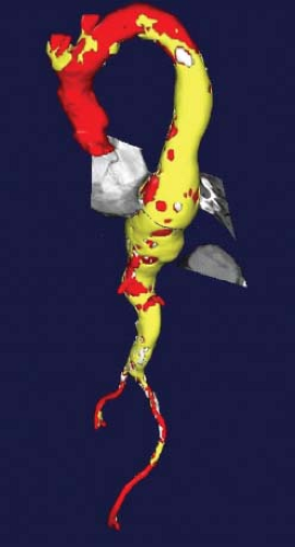
Figure sixteen.3 Iii-dimensional reconstruction of the thoracoabdominal aorta with an centric window in the centerline plane allows for the nigh accurate measurement of aneurysm diameter.
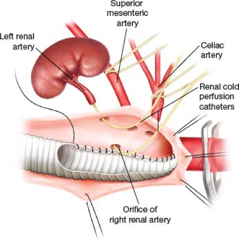
Figure sixteen.four Askew proximal anastomosis includes the right renal artery, SMA, and celiac. Renal cold perfusion catheters are in both renal arteries. The posterior suture line is performed starting time.
Positioning
Irrespective of individual preferences concerning the technical components of the operation, the key to operative success remains the provision of broad, continuous exposure of the entire left posterolateral aspect of the thoracoabdominal aorta. The patient is positioned on the table in the right lateral decubitus position. The location and extent of the thoracic portion of the incision is adamant by the proximal extent of the aneurysm. A thoracoabdominal incision at the eighth interspace volition ordinarily provide adequate exposure for a Blazon Iv aneurysm, and a double lumen tube for deflation of the left lung is mostly non necessary in these cases (Fig. 16.5
But gold members can continue reading. Log In or Register to continue

What Type Of Aortic Anuerysm Is Easiest To Repair,
Source: https://thoracickey.com/type-iv-thoracoabdominal-aneurysm-repair/
Posted by: gonsalvesextres47.blogspot.com
0 Response to "What Type Of Aortic Anuerysm Is Easiest To Repair"
Post a Comment